ID photo software
Сamera control software
ID card software

info@akond.net, akondsales@gmail.com

  User stories >> Fungi microscopy
 
Home
News
Products
ID Photo Solutions
Download
Buy
Support
Applications
Company
Links
Contact
 
Fungi microscopy


inPhoto Capture application:
Fungi microscopy


This story is the courtesy of Mr. Fermin Pancorbo, Product Engineering Manager, Indra Sistemas, Madrid, Spain. Mr. Fermin Pancorbo won a free copy of inPhoto Capture camera control software designed for full computer control of Canon and Olympus digital cameras). Please refer to giveaway action for more details.


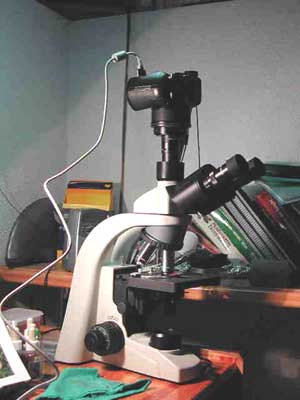
Figure 1. Imaging system (Olympus C-8080WZ camera
based on Motic BA 200 microscope).
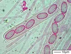
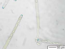
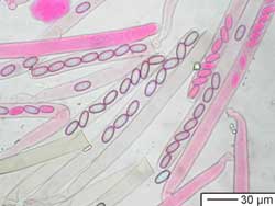
In order to document my fungi findings, I use to write down a record where annotate all the outstanding data as place, date, habitat, UTM co-ordinates, pictures of the mushroom in their habitat and later on, at home, I carry out a microscopic analysis to complete the record.

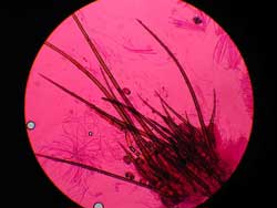
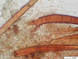
Taking photographs of the microscopic structures such as spores, cystidia, hyphae, basidia, etc is for me a must because I use these photos to measure this structures with a special measurement software, so it is important to get a sharp and clear image.

The configuration of the imaging system I use is as follows:

Camera: Olympus C-8080WZ,
Microscope: Motic BA 200,
Adapters rings from 58 to 28mm,
28mm Leitz Periplan 10x18 Objective.


With a Compact Digital Camera it is not easy to focus properly just looking to LCD Camera screen. So I have been looking for a capture software with a live preview.

Another important feature is the ability of taking several photos in a remote mode varying slightly the microscope focus over the same object in order to assemble them and obtain an image with more "deep of field" like a 3D image.

I found several programs and I have been working in a trial mode with them. With InPhoto Capture I have these features and it is the best remote capture software that I have tested until now.
 
Figure 2. Peziza echinospora spores X1000.
Figure 3. Peziza echinospora tip asci amyloid X400.
 
Figure 4. Peziza echinospora asci X400.
 
Figure 5. Scutellinia crinita hairs X100.
 
Figure 6. Scutellinia crinita hairs. Detail of base X400.


The main problem with the contact endoscopy is their reliability and difficulties in diagnostic interpretation with standard PAL-video signal which is commonly used in standard.


When we acquire images of microalgae from a compound microscope it is very important to be able to adjust focus correctly to shoot a sharp image useful for scientific publications, and with inPhoto Oly software we now have this ability, which saves us much time and effort. We look forward to sharing the beauty and secrets of tropical marine algae with these new technological tools now at our disposal.


  See also:
 
Home|News|Products|ID Photo Solutions|Download|Buy|Support|Applications|Company|Links|Contact


Software Informer
Contact: AKOND company
Tel: +7-921-7578319
Skype: alla.povarkova
email: info@akond.net
email: akondsales@gmail.com

Copyright © 2006 - 2015, Akond
Web site designed and developed by Memento Graphics